


It seems that nicer weather equates to an increase in hamstring injuries!
This past week, I had three cases of hamstring strain present in the clinic, all with a similar history of increasing training intensity as a result of pushing it harder now that the weather allows for outside training.
Previously, Absolute has discussed hamstring injuries in detail in previous posts as well as in a Founders Meeting discussing the New Ecology of the Hamstrings.
In addition, we have previously presented the Internal Isometric Continuum (IIC) and how the use of varying isometric inputs within different timeframes from an initial injury presentation to return to performance is a very effective approach to cultivating differing strength behaviours along the progression of tissue healing, remodelling and normalization.
Using one of the cases of an acute hamstring injury as an example, as well as the IIC we wanted to demonstrate the initial management of this type of soft tissue based injury and its effects on the initial behaviours of strength as governed by the neural network.
Case: Information on Point A

One of the clients from this past week, is a male 100/200M sprinter, with no history of previous hamstring injury who recently increased his training volume adding more speed endurance work to his regimen, adding tempo work more times per week at 70% effort for repeated bouts of 8-10. This was the major difference in training over the last few weeks.
The injury occurred towards the end of a recent training session. The athlete reports that on initial ground contact he felt the hamstring “give” and felt an immediate concentration of pain located in the proximal medial side. He was able to bear weight on the leg but was unable to fully move the hip and extend the knee during walking after the injury. On subsequent days he felt an area of extreme tightness in the upper hamstring with no visible bruising. He reported to the clinic 5 days after injury. The timing of the injury is important. According to the literature this is a common time during the sprint cycle when injury is likely to occur on the medial side as the leg is absorbing impact forces while also controlling for hip extension under forward momentum and stabilizing the knee by controlling tibial internal rotation.
In this athlete’s case it may seem obvious that a specific increase in demand without adequate tissue preparation for increased distance and volume would change the web of determinants in such a way as to increase the potential for a subsequent hamstring injury. As this may be the case the timing of the injury during the sprint cycle, as well as the location of the injury (medial side) suggest clues as to the capacities that may need to be trained to move this athlete to Point B.
On examination, using Functional Palpation1 the athlete reports pain with perpendicular palpation in the proximal medial hamstring (semimembranosus) and in the epimysial interface between the medial and lateral hamstrings. Specific tissue assessment using the length and tension properties of the tissue was not performed as the hamstring was unable to be moved at length as a result of underlying spasm.

Using Functional Range Assessment,2 the passive range of motion of the hip was assessed for the fundamental joint motions. On the affected side internal rotation was limited when compared to the opposite side. No other hip motions were assessed.
Ratios of knee range of motion were assessed and found to be full passively but with a limitation in active knee flexion on the shorter side of 90 degrees.
Considerations for Point B
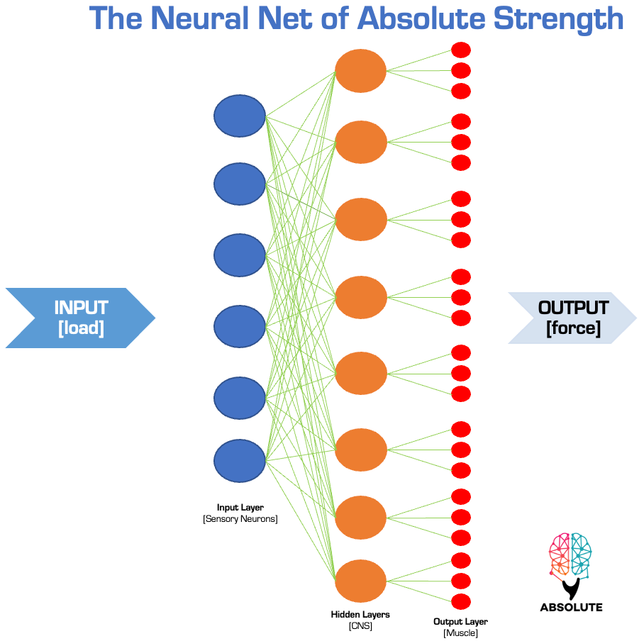
Although all physical capacities associated with Point B will need to be considered and effectively trained during the program to return to sprinting, at this stage so early in the management it is paramount that the effects of spasm are minimized and the effects of neural network compromise are addressed as this is the current main physical limiting factor and will have the potential to create motor output deficiencies later on in the program. The neural network is the governor to all other strength capacities, so therefore becomes a priority to repeatedly stimulate at this stage.
Spasm and its Effects on the Neural Network
After soft tissue injury occurs a protective spasm in the muscle associated. A spasm is a sudden, involuntary reflexive contraction of a muscle or group of muscles. Physiologically, a spasm occurs due to a complex interplay between the nervous system and the muscle fibers. There are other potential factors at play such as local tissue ischemia, electrolyte imbalances as well as nutrient deficiencies, however for ease of explanation only the factors that involve the neural net locally and globally will be discussed.
Local Effects
Locally within the injured tissue will be a very fast acting inflammatory response that is initiated by a variety of biochemical mediators (prostaglandins, interleukins, etc.) that will create sensitization of sensory nerve endings in the local area of the injured muscle. Ultimately this creates altered afferent information to the CNS. This creates a large amount of noise within the nervous system and alters the efferent output creating a reflexive increase in muscle tone triggering muscle spasm.
At the same time, due to the sustained output of the nervous system there exists increased tension on the muscle spindles promoting the neurophysiological effects of the myotactic reflex furthering triggering the spasm leading as a result to sustained contraction evident by actin/myosin overlap. This leads to excessive chemical build-up within the muscle leading to a closed loop of sustained spasm which for the short term has necessary protective effects, but in the slightly longer term has a direct impact on the recovery process.
Global Effects
The more global effects of acute muscle spasm occur within the spinal cord and cortex and have both a direct and indirect effect on the descending modulation of pain and muscle activity. Normally, these descending effects, referred to as descending inhibition are aimed at modulating noisy neural signals and suppressing excessive muscle activity related to the acute spasm. One of the major components of descending inhibition is the reaction of the motor cortex to send inhibitory signals to the lower CNS to regulate excessive muscle contractions of the injured tissue. In turn this creates the activation of inhibitory interneurons within the anterior horn of the spinal cord to minimize the polarization of alpha motor neurons effectively shutting down the output mechanism for muscle activation. Although this has well-meaning implications, the prolonged effects of minimizing the motor neuron pool can have implications for muscle force potential in the later phases of training.
Using the Internal Isometric Continuum to Influence the Neural Network
Keep reading with a 7-day free trial
Subscribe to Absolute: The Art and Science of Human Performance to keep reading this post and get 7 days of free access to the full post archives.