Training an Adductor Strain Injury: An Alternative Strategy to the Copenhagen
A case study of treatment and training strategy for the adductor tissues.
In a prior article focused on adductor tissue training, we delved into the scientific literature to articulate our contention with the prevailing consensus that the Copenhagen training strategy effectively rehabilitates connective tissue injuries in the adductors. In the same manner, we have shared our views on the limitations of the Nordic hamstrings curl as the training solution for the widespread epidemic of hamstring injuries across all levels of athletes in modern sports (see: The Hamstrings: A New Ecology). Rather than reiterating this stance, as we've made it clear, our objective is to introduce a logical treatment and training strategy for the athlete who currently has sustained an adductor injury.
Adductor Anatomy
The medial thigh region, although straightforward from a muscle perspective, is actually quite complex when considered within an anatomical continuum as well as the function of that continuum.
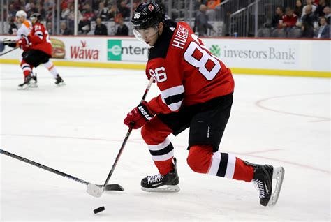
The first consideration of the anatomy is the linkage of the adductor region with the trunk of the opposite side, specifically the external oblique, serratus anterior, latissimus, and pectorals major, which all have finer orientations in the same direction as the adductor group. From a skating perspective, this linkage is important as the leg strides outward and backward, the ipsilateral arm drives forward, loading the opposite side trunk, building a large amount of potential energy to be returned during the next stride. This connection of the trunk to the opposite hip and its role in energy transfers during locomotion should be considered in any adductor training (rehabilitation) program.
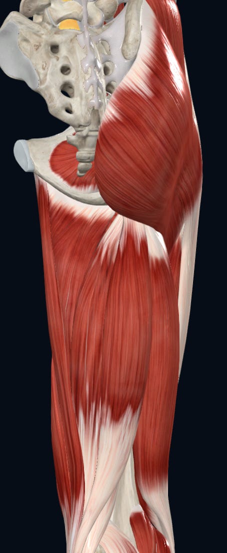
The second consideration with respect to the adductor region is the anatomy of the adductors themselves (brevis, longus, magnus). These tissues all begin proximally on differing locations of the pubic bone and run in medial superior to lateral inferior direction onto the femur, creating a line of action whereby the femur is pulled to the midline. However, it is not as simple as this in reality. As a result of this anatomical configuration, each adductor will have two sides: an anterior and a posterior, often referred to as "faces" in Functional Range Systems.


As you can see from the anatomical slides above, there is depth to the adductors that is often not considered in the traditional view of what constitutes the medial thigh and its training. Functionally, these faces are important for the complex movements of the hip, particularly with reference to the skating motion as done by the athlete in our case. Simply put, the two faces of each adductor will have different behavioural demands simultaneously. For example, during adduction and extension of the femur, the anterior faces will be lengthened under load (eccentrically), while the posterior face will shorten under load end range. On stride return, the opposite will occur. It also must be considered that in the athletic position, the two faces will be under differing loads from the outset; subsequent movement will determine further dynamic loading.
Case Study
To provide some specifics on the initial injury, it occurred during the course of a hockey game, whereby the right leg was extended out in a stride and slid excessively on an opposing player’s stick. Imaging was sought and showed connective tissue disruption on the proximal adductor longus near the pubic bone.
Physical Assessment of the Athlete:
The athlete presented with pronounced neurological tightness within the entire lower extremity. On the passive assessment of the fundamental ranges of motion of the hip (i.e., internal and external rotation), there was a distinctly bouncy neurological barrier with no stretching sensation within the hip joint capsule on behalf of the athlete. Notably, there was no closing angle block or pain at the end range of motion, only a bouncy neurological barrier with no capsular stretching sensation on the athlete’s or practitioner’s behalf.
The heightened neurological tightness significantly limited the passive range of motion of the hip joint. Despite conducting multiple PAILs tests, with each repetition leading to a noticeable and immediate decrease in neurological tightness, there was still no stretching sensation within the connective tissue architecture of the hip joint capsule.
During the passive range of motion assessment, the athlete expressed apprehension, verbally asking if we were going to make the tear worse through passive assessment. It was communicated to him that elevated neurological tightness was so high that we were not able to actually assess the behavior of the connective tissue architecture (i.e., the length-tension relationship).
In conjunction with the questions of making the tear worse, the apprehension signs showed two neurological tightness positive feedback loops reinforcing each other - one local at the hip level and one at the level of consciousness. These feedback loops had been reinforced for so long that even multiple PAIL tests did not lower them to the point where we could accurately assess the behavior of the hip joint capsule or the connective tissue architecture of the adductor longus.
The findings from the physical assessment enabled us to know that we had to specifically deal with the neurological tightness before we were able to apply force-based input into the connective tissue architecture. More simply, the nervous system of this athlete was acting as the gatekeeper, controlling access to the injured connective tissue. The subsequent protocol for this specific case (detailed below) highlights the extensive use of electrical muscle stimulation (EMS), which performed conjugate in the clinic with PAILs proved successful in rapidly reducing the neurological tightness barrier. This allowed us to assess the connective tissue behavior in the hip joint capsule and adductor longus and apply force-based inputs to specific tissues that exhibited abnormal behavior.
We will delve more into the specifics of this injury in future videos (any questions you have, please leave in the comments section), but for the context of understanding the training strategy we are presenting, we want subscribers to understand the Copenhagen training strategy failed this athlete. The injury occurred over seven weeks ago, and at that point in time, an MRI was performed. After 5-6 weeks of performing the Copenhagen eccentrics, the connective tissue tear worsened - exactly what we wrote as a real possibility of occurring.
So this athlete was seven weeks post-acute injury, and their connective tissue architecture is in a worse physical state - due to the blindspot that connective tissue architecture is a trainable biological element of strength and illogical utilization of the Copenhagen.
Absolute’s Alternative to the Copenhagen
Keep reading with a 7-day free trial
Subscribe to Absolute: The Art and Science of Human Performance to keep reading this post and get 7 days of free access to the full post archives.


